

What is Magnetic Resonance Imaging?
Magnetic Resonance Imaging (MRI) is a modern and safe diagnostic procedure that enables detailed visualization of the body’s interior. It uses a high-energy electromagnetic field to produce detailed images of soft tissues, organs, and the nervous system. MRI is particularly valuable in the diagnosis of neurological, orthopedic, and oncological conditions.
This examination is safe because, unlike computed tomography (CT), it does not use ionizing radiation. The procedure is usually painless, although some patients may experience discomfort due to the confined space of the MRI tunnel. Prior to undergoing an MRI scan, patients must disclose any contraindications, such as metallic implants in the body.
MRI at Rehasport
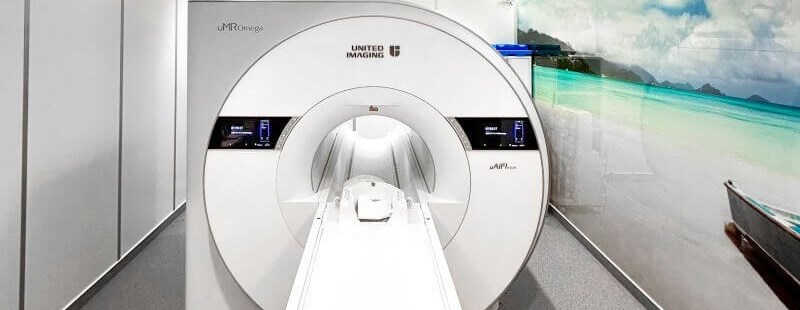
One of the most advanced MRI facilities in Poland is located on the ground floor of the Panorama Center at 30 Górecka Street in Poznań. Rehasport is equipped with a state-of-the-art 3T MRI scanner manufactured by United Imaging, featuring the latest global technological and technical innovations.
The uMR Omega™ model utilizes uAIFI technology, significantly enhancing performance and expanding imaging capabilities. This system provides high-quality images without prolonging scan times, which greatly improves patient comfort.
The 3T MRI scanner used at Rehasport is the world’s first with a 75 cm wide bore, offering a unique, starry-sky-like environment to ensure patients feel as comfortable as possible during the examination.
What Does an MRI Scan Involve?
MRI involves the use of a strong magnetic field and radio waves to generate detailed images of internal structures. Depending on the area being examined, the patient lies either on their back or stomach on a special motorized table, which slides into the tunnel-shaped scanner. During the scan, the patient must remain still, as any movement may affect image quality.
MRI is painless, but the scanner can produce loud noises during the procedure. Therefore, patients are often given earplugs or headphones. Throughout the exam, the patient is monitored by medical personnel and can communicate via a microphone, camera, or emergency button.
The entire scan typically takes between 15 and 60 minutes, depending on the area being assessed. In some cases, a contrast agent is administered intravenously to enhance visualization of certain tissues.
What is Contrast-Enhanced MRI?
Contrast-enhanced MRI is a special type of scan in which a contrast agent—most commonly gadolinium-based—is administered intravenously before or during the procedure. The contrast helps to better visualize certain anatomical structures, enabling a more accurate diagnosis.
This substance increases the contrast in the way tissues reflect radio waves, making abnormal or diseased areas more distinguishable on the images. Contrast-enhanced MRI is particularly helpful in diagnosing tumors, neurological disorders, and cardiovascular diseases.
The contrast agent is usually well-tolerated and painless to administer, although some patients may feel a temporary sensation of warmth. Prior to undergoing contrast-enhanced MRI, a physician will assess the patient's health, especially kidney function, as the contrast medium may place stress on the kidneys.
Indications for MRI
MRI is recommended in many situations where detailed imaging of internal structures is required, particularly soft tissues that are difficult to visualize using other techniques.
The most common indications include:
- Neurological conditions such as multiple sclerosis, brain tumors, strokes, or meningitis
- Spinal disorders such as disc herniation, degenerative disc disease, and spinal cord injuries
- Musculoskeletal conditions affecting joints, ligaments, muscles, or surrounding tissues (especially useful in orthopedics)
- Oncological diagnostics—for tumor detection and staging
- Vascular abnormalities such as aneurysms, thrombosis, or congenital vessel malformations
Advantages of MRI at Rehasport
- Higher precision due to the latest imaging technology
- More accurate diagnosis thanks to the clinical specialization of the facility and medical team
- Greater patient comfort with all essential imaging techniques centralized in one location
- Comprehensive care with responsibility for the full treatment process—from diagnostics and consultation to surgery and rehabilitation
- Short waiting times for appointments
How to Prepare for an MRI Scan
- Wear clothing free of any metallic elements, or be prepared to remove them before the scan.
- Bring results of any previous imaging exams (X-ray, ultrasound, CT, MRI).
- Leave your phone, MP3 player, and other electronic devices in the locker room—they will be safe there.
- Remove watches, jewelry, belts, and any other items containing metal before the scan.
- If you wear contact lenses, be ready to remove them if undergoing a head scan.
- If the head is being scanned, avoid wearing makeup or remove it beforehand (cosmetics may contain trace amounts of metal).
- Fasting is not necessary unless an abdominal scan is scheduled.
Contraindications to MRI
- Metal orthodontic braces
- Artificial heart valves
- Implanted pacemaker
- Cochlear (middle ear) implants
- Retained metallic foreign bodies
- Vascular clips unless certified as non-metallic by the manufacturer
- Pregnancy
- Foreign bodies such as joint prostheses, bone fixations, or tracheotomy tubes

